Review sheet 11 microscopic anatomy and organization of skeletal muscle – Unveiling the intricate world of skeletal muscle, this review sheet delves into the microscopic anatomy and organization that governs its remarkable contractile properties. From the structure of individual muscle fibers to the arrangement of fascicles within a muscle, we embark on a journey to unravel the fundamental principles that underpin skeletal muscle function.
This review sheet provides a comprehensive overview of the microscopic anatomy of skeletal muscle, exploring the structure and organization of muscle fibers, myofilaments, and sarcomeres. It also examines the role of connective tissue in organizing muscle fibers into fascicles and muscles, and discusses the functional implications of muscle structure on its contractile properties.
Overview of Skeletal Muscle
Skeletal muscle, also known as voluntary muscle, is a type of muscle tissue that is attached to bones and enables voluntary movement. It plays a crucial role in locomotion, posture maintenance, and other voluntary actions.
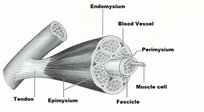
Skeletal muscle tissue is organized into individual muscle fibers, which are long, cylindrical cells that contain specialized contractile proteins. These muscle fibers are grouped into bundles called fascicles, which are further enveloped by connective tissue layers to form a complete muscle.
Microscopic Anatomy of Skeletal Muscle

Structure of a Muscle Fiber
Each muscle fiber is surrounded by a plasma membrane called the sarcolemma. The cytoplasm of the muscle fiber, known as the sarcoplasm, contains numerous myofibrils, which are composed of actin and myosin filaments.
Actin filaments are thin and contain a binding site for myosin heads, while myosin filaments are thick and have two globular heads that can bind to actin.
Arrangement of Myofilaments
Myofilaments are arranged in a repeating pattern called a sarcomere, which is the basic unit of muscle contraction. Sarcomeres are defined by Z-lines, which are dense protein structures that anchor actin filaments from adjacent sarcomeres.
Within a sarcomere, the I-band contains only actin filaments, the A-band contains both actin and myosin filaments, and the H-zone is the region of the A-band that contains only myosin filaments.
Organization of Skeletal Muscle

Muscle Fiber Organization
Muscle fibers are organized into fascicles, which are bundles of parallel fibers surrounded by a layer of connective tissue called the perimysium.
Fascicles are further grouped into a complete muscle, which is enveloped by a tough connective tissue layer called the epimysium.
Connective Tissue
Connective tissue plays a crucial role in the organization and function of skeletal muscle. The endomysium surrounds individual muscle fibers, providing structural support and facilitating nutrient and waste exchange.
The perimysium surrounds fascicles, providing strength and stability to the muscle. The epimysium envelops the entire muscle, protecting it from damage and providing attachment points for tendons.
Functional Implications of Muscle Structure

Relationship between Structure and Contraction, Review sheet 11 microscopic anatomy and organization of skeletal muscle
The structure of skeletal muscle is directly related to its contractile properties. The arrangement of myofilaments in sarcomeres allows for efficient force generation and muscle shortening during contraction.
The presence of sarcoplasmic reticulum and transverse tubules ensures rapid and coordinated muscle contraction.
Role of Sarcoplasmic Reticulum and Transverse Tubules
The sarcoplasmic reticulum is a network of membranes that stores calcium ions. When a nerve impulse reaches the muscle fiber, it triggers the release of calcium ions from the sarcoplasmic reticulum, which bind to receptors on the transverse tubules.
Transverse tubules are invaginations of the sarcolemma that conduct the nerve impulse deep into the muscle fiber, allowing for rapid and uniform muscle contraction.
Neuromuscular Junction
The neuromuscular junction is the site where a motor neuron communicates with a muscle fiber. When an action potential reaches the nerve terminal, it triggers the release of neurotransmitters, such as acetylcholine, which bind to receptors on the muscle fiber, leading to muscle activation.
Essential Questionnaire: Review Sheet 11 Microscopic Anatomy And Organization Of Skeletal Muscle
What is the role of the sarcoplasmic reticulum in muscle contraction?
The sarcoplasmic reticulum stores calcium ions, which are released during muscle contraction to trigger the interaction between actin and myosin filaments.
How does the neuromuscular junction contribute to muscle activation?
The neuromuscular junction is the site where motor neurons communicate with muscle fibers. When a motor neuron fires an action potential, it releases neurotransmitters that bind to receptors on the muscle fiber, triggering muscle contraction.